10.3 Mechanism of Heartbeat
The heart functions as a muscular pump that continuously circulates blood throughout the body. Unlike ordinary muscles, cardiac muscles are interconnected and myogenic, meaning they possess the innate ability to contract and relax rhythmically without needing nerve impulses from the central nervous system. This topic explores how electrical impulses are generated by internal pacemakers, how they coordinate the rhythmic pumping of atria and ventricles, the origin of heart sounds, and how blood returns to the heart against gravity.
Learning Objectives
- Define the term myogenic in relation to cardiac muscles.
- Identify the key structures of the electrical conduction system in the heart: sinoatrial (SA) node, atrioventricular (AV) node, bundle of His, and Purkinje fibres.
- Describe the sequence of electrical impulse generation and conduction that coordinates atrial and ventricular contractions.
- State the causes of the "lub-dub" heart sounds.
- Explain the mechanisms assisting venous blood return to the heart, including skeletal muscle contraction and venous valves.
The Electrical Conduction System and Heartbeat Sequence
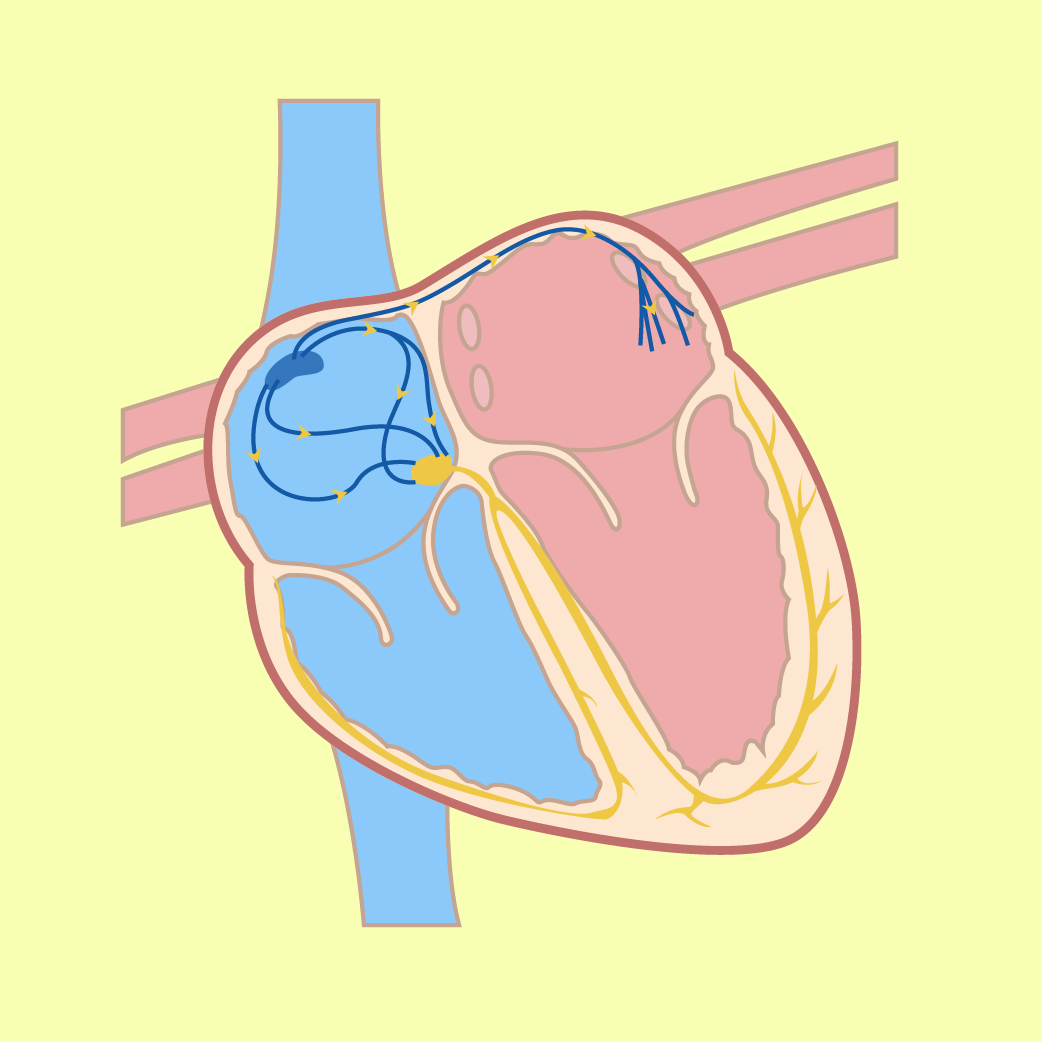
A clear spatial and chronological view of how an impulse travels from the SA node to the Purkinje fibres, accompanied by valve actions causing the "lub-dub" sound.
1. SA node
The sinoatrial (SA) node generates electrical impulses.
2. Atrial contraction
Impulses spread rapidly through both atria walls, causing both atria to contract simultaneously and pump blood into the ventricles.
3. AV node → apex
Impulses reach the atrioventricular (AV) node, then travel down through the bundle of His and Purkinje fibres to the apex of the heart.
4. Ventricular contraction
Impulses spread upwards from the apex through the entire ventricle walls, causing both ventricles to contract simultaneously to pump blood out to the lungs and the rest of the body.
"Lub" — First Heart Sound
Produced when the atrioventricular valves (tricuspid valve and bicuspid valve) snap shut during ventricular contraction.
"Dub" — Second Heart Sound
Produced when the semilunar valves (aortic and pulmonary semilunar valves) snap shut during ventricular relaxation.
Short Explanation
Myogenic Cardiac Muscle
Myogenic means that cardiac muscles contract and relax without receiving any impulse signal from the nervous system. Cardiac muscle cells intersect and connect with one another, allowing electrical impulses to spread rapidly and stimulate cells to contract simultaneously and uniformly. If cardiac muscles are maintained in a warm, oxygenated nutrient solution, they will continue to contract and relax rhythmically on their own.
Pacemaker
A pacemaker is a specialized group of cardiac muscle cells located in the right atrium wall that initiates and sets the rate of heart contractions. The sinoatrial (SA) node is known as the main pacemaker of the heart.
Electrocardiogram (ECG)
An electrocardiogram (ECG) is a clinical record of the electrical activity that triggers each heartbeat, measured via electrodes placed on the skin.
Artificial Pacemaker
An artificial pacemaker is a miniature device, such as the "Medtronic Micra" — the size of a vitamin pill — placed in the heart without major surgery to send small electrical charges to stimulate heartbeats when natural regulation fails.
Blood Circulation Forces
Blood circulation is driven by two main forces: the pumping force generated by the heart, and the contraction of skeletal muscles around the veins.
Try to Answer First
Answer in your mind, then press "Check Answer".
What does the term "myogenic" mean in relation to cardiac muscles?
What causes the first heart sound ("lub")?
Why does blood inside the veins of the lower limbs continue to move towards the heart against gravity?
Common Mistakes
- Assuming that every heartbeat is triggered directly by a signal from the brain or nervous system, ignoring the intrinsic myogenic property and the role of the SA node.
- Confusing the valves responsible for each heart sound — attributing "lub" to semilunar valves and "dub" to atrioventricular valves instead of the reverse.
- Believing that heart sounds are caused by blood rushing through the chambers rather than the sudden closing of heart valves.
- Thinking that the pumping force of the heart alone is strong enough to push venous blood from the lower legs back to the right atrium without skeletal muscle assistance.
Concept Misunderstandings
The heart stops immediately if it is disconnected from the nervous system.
Because cardiac muscle is myogenic, an isolated heart placed in an oxygenated, warm nutrient solution will continue to contract and relax rhythmically on its own.
Valves produce sounds when they open to let blood pass.
Heart sounds ("lub" and "dub") are exclusively caused by the snapping closure of the valves, preventing backflow.
Summary
- Cardiac muscles are myogenic: they initiate their own contractions without nervous stimulation.
- The sinoatrial (SA) node in the right atrium wall is the main natural pacemaker.
- Impulses spread from the SA node → atria walls (atrial systole) → AV node → bundle of His → Purkinje fibres → ventricular walls from the apex (ventricular systole).
- "Lub" sound = closure of tricuspid and bicuspid valves.
- "Dub" sound = closure of semilunar valves.
- Venous return against gravity is powered by the contraction of skeletal muscles compressing veins, coupled with one-way valves that prevent backflow.
Short Activity
Objective: test your understanding of the heartbeat mechanism, heart sounds and venous return.
A. Objective Quiz
Which structure serves as the primary natural pacemaker that initiates the rate of heartbeat in the human heart?
The second heart sound, "dub", is produced when which of the following events occurs?
B. Fill in the Blanks
Cardiac muscle is described as because it contracts and relaxes rhythmically without receiving impulse signals from the nervous system.
The first "lub" sound of the heartbeat is produced when the semilunar valves remain open and the tricuspid and valves snap shut.
C. Matching / Drag and Drop
Match each component of the cardiovascular conduction and venous system to its correct description. On a phone, tap a choice first, then tap the matching box. Tap the Answer Choices area to return a selected item.
C. Match the Component to Its Function
Write the correct component next to each description.
| Function / Description | Component |
|---|---|
| 1. Main pacemaker located in the right atrium wall that generates electrical impulses | |
| 2. Specialised fibres that spread impulses from the apex through the entire ventricular wall | |
| 3. Structures inside veins that close to prevent backflow of blood towards the lower limbs | |
| 4. Heart valves whose closure produces the second ("dub") heart sound |
Answer choices: Sinoatrial (SA) node • Purkinje fibres • Venous valves • Semilunar valves