10.2 Circulatory System of Humans
The human circulatory system is a vital transport network consisting of three main components: blood, the heart, and blood vessels. Blood acts as a circulating transport medium, the heart functions as a muscular pump to generate pressure, and blood vessels form a closed circuit of tubes that convey blood to every tissue throughout the body.
Learning Objectives
- Identify the three main components of the human circulatory system.
- Describe the internal and external structure of the human heart and the functions of its chambers, valves, and associated blood vessels.
- Explain why the muscular wall of the left ventricle is thicker than that of the right ventricle.
- List the components of human blood and outline the main functions of blood plasma, erythrocytes, platelets, and leucocytes.
- Differentiate between granulocytes (neutrophils, eosinophils, basophils) and agranulocytes (lymphocytes, monocytes).
- Compare and contrast the structural characteristics and functions of arteries, capillaries, and veins.
Internal Anatomy of the Human Heart and Blood Vessel Structural Comparison
The heart is a dual pump with four chambers and valves that keep blood flowing in one direction. Blood vessels differ in wall thickness, lumen size, and the presence of valves.
Blood
A connective tissue made up of blood plasma, blood cells, and platelets that functions as a transport medium.
Heart
A muscular pump located in the thorax cavity between the lungs, approximately the size of a fist, which circulates blood to the entire body.
Blood Vessels
A network consisting of arteries, capillaries, and veins connected to the heart to carry blood to all body tissues.
Short Explanation
Main Components of the Circulatory System
The human circulatory system consists of blood, the heart, and blood vessels. Coronary arteries deliver oxygenated blood to cardiac muscle tissues, while coronary veins transport deoxygenated blood away from the heart tissues.
Structure of the Human Heart
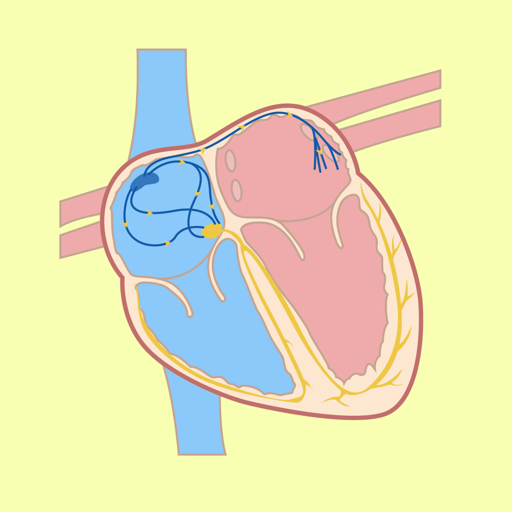
The heart has four chambers: thin-walled atria that receive blood, and thick-walled ventricles that pump blood out. The septum separates the left and right sides. The tricuspid valve lies between the right atrium and right ventricle, while the bicuspid valve lies between the left atrium and left ventricle. Semilunar valves are situated at the base of the aorta and pulmonary artery.
Why is the left ventricle thicker?
The muscular wall of the left ventricle is much thicker than that of the right ventricle. The left ventricle must generate greater pressure to pump oxygenated blood through the aorta to the entire body, whereas the right ventricle only pumps deoxygenated blood a short distance to the lungs.
Composition of Human Blood
Blood is composed of 55% blood plasma and 45% cellular components (erythrocytes, leucocytes, and platelets). Plasma contains water, plasma proteins such as fibrinogen, albumin and globulin, nutrients, excretory substances, respiratory gases, hormones and enzymes.
Cellular Components of Blood
Erythrocytes transport oxygen. Platelets are essential for blood clotting. Leucocytes defend the body against pathogens and are divided into granulocytes and agranulocytes.
Try to Answer First
Answer in your mind, then press “Check Answer”.
Why is the muscular wall of the left ventricle thicker than that of the right ventricle?
State two structural adaptations of a mature human erythrocyte and their respective advantages.
What is the functional importance of valves in human veins?
Common Mistakes
- Confusing the positions of the atrioventricular valves: misidentifying the bicuspid valve as being on the right side and the tricuspid valve on the left side. Remember: Tricuspid is on the Right, Bicuspid is on the Left.
- Stating that arteries carry oxygenated blood and veins carry deoxygenated blood without noting exceptions: the pulmonary artery carries deoxygenated blood to the lungs, while pulmonary veins carry oxygenated blood to the heart.
- Believing that arteries have valves along their length: arteries do not have valves along their tubes; semilunar valves are only present at the base of the aorta and pulmonary artery.
- Stating that mature erythrocytes lack a nucleus because they are dead cells: erythrocytes lack a nucleus at maturity specifically to accommodate more haemoglobin molecules.
Concept Misunderstandings
Blood in veins flows backwards whenever a person stands up.
Veins contain pocket valves that close immediately when blood attempts to flow backwards, ensuring blood flows in one direction toward the heart.
The septum is just a dividing line between chambers without physiological significance.
The septum is a thick muscular wall that completely separates the left and right sides of the heart to prevent oxygenated blood from mixing with deoxygenated blood, maintaining high oxygen delivery efficiency.
Blood capillaries have thin walls made of muscle tissue.
Capillary walls contain no muscle or elastic fibers; they consist solely of a single layer of endothelial cells to minimise diffusion distance.
Summary
- The human circulatory system consists of the heart, blood vessels, and blood.
- The heart contains four chambers: right atrium, right ventricle, left atrium, and left ventricle.
- The left ventricle has the thickest muscular wall to pump blood to the entire systemic circulation.
- Valves (tricuspid, bicuspid, semilunar) maintain unidirectional blood flow.
- Blood plasma (55%) transports water, proteins (fibrinogen, albumin, globulin), nutrients, and wastes.
- Cellular components (45%) comprise erythrocytes (oxygen transport), platelets (blood clotting), and leucocytes (body defence).
- Leucocytes are divided into granulocytes (neutrophils, eosinophils, basophils) and agranulocytes (lymphocytes, monocytes).
- Arteries carry blood away from the heart under high pressure; capillaries allow diffusion exchange; veins return blood to the heart under low pressure with the aid of valves.
Short Activity
Objective: check your understanding of the heart, blood components, and blood vessels.
A. Objective Quiz
Which of the following statements correctly explains why the wall of the left ventricle is thicker than the wall of the right ventricle?
Which leucocyte contains heparin that prevents blood clotting?
B. Fill in the Blanks
The muscular wall that separates the left chamber of the heart from the right chamber to prevent the mixing of oxygenated and deoxygenated blood is called the .
The erythrocyte has a disc shape which provides a large total surface area to volume ratio for efficient gaseous exchange.
C. Matching / Drag and Drop
Match each blood cell component with its correct function or description. On a phone, tap a choice first, then tap the matching box. Tap the Answer Choices area to return a selected item.
C. Match the Blood Cell Component to Its Function
Write the correct blood cell component next to each function.
| Function or Description | Blood Cell Component |
|---|---|
| 1. Ingests bacteria and dead cells from wounds by phagocytosis | |
| 2. Produces antibodies and antitoxins to destroy pathogens | |
| 3. Cell cytoplasm fragments involved in the blood clotting process | |
| 4. Releases enzymes that fight inflammation and allergy reactions |
Answer choices: Neutrophil • Lymphocyte • Platelet • Eosinophil